Have you wondered about the process of an autopsy, when it may be required, and considerations if you request one? We answer all these questions and more in this episode.
When is an Autopsy Required?
If the death is sudden and unexpected or if it occurs at home, the medical examiner must be notified, and he or she will decide if an autopsy is required. The family has no authority to stop an officially mandated autopsy.
The next of kin may request an autopsy even if the medical examiner declines to do one. An autopsy will help determine the cause of death, but the family will be charged a fee for this service. Autopsies are not covered under Medicare, Medicaid, or most insurance plans, though some hospitals — teaching hospitals in particular — do not charge for autopsies of individuals who died in the facility. A private autopsy by an outside expert can cost between $3,000 and $5,000. In some cases, there may be an additional charge for the transportation of the body to and from the autopsy facility.
Autopsies are best if performed within 24 hours of death, before organs deteriorate, and ideally before embalming, which can interfere with toxicology and blood cultures. But autopsies performed on decomposed or even exhumed bodies can still provide vital new information, depending on the extent of decomposition. An autopsy will generally take two to three hours to complete, except if there are excessive complications.
Autopsies also serve other purposes:
| The Uses of Autopsies |
|---|
| Benefits to Medical Practice and Science Discover or clarify new diseases Explain unknown or unanticipated medical complications Assist in the development/quality assurance of new technology, procedures, and therapy Educate medical students Continue physician education |
| Benefits to the Judicial System Classify and explain sudden, unexpected, and/or unnatural deaths |
| Benefits to Public Welfare Identify infectious and contagious diseases Identify and monitor occupational and environmental health hazards Quality control and risk assessment in hospital practices Provide a source of organs and tissues for medical and scientific purposes Provide materials and hypotheses for research Improve accuracy, and therefore usefulness, of vital statistics |
| Benefits to the Deceased’s Family Assist in the grief process Provide a vehicle for contribution Discover contagious diseases within the family Assist in genetic counseling and identification of family health risks Provide information for insurance/death benefits |
Autopsies and Organ Donation
If the deceased has requested that his or her organs be donated, the nurse is often the person responsible for notifying the proper agencies for organ and tissue harvesting. Organ donation is the practice of giving a part of the deceased body for transplantation into another person. Persons designate this wish to donate organs by signing the back of their driver’s license, indicating their preferences; by specifying organ donation in an advance directive; or by filling out an organ donor card. Listen to our podcast about organ donation for more information.
Depending on what organ is being donated, the time for organ removal varies. Eyes must be removed within 4 hours of the death but once they are in a preservative, they can wait 10 to 14 days to be transplanted. Tissue (e.g., bone, skin, and tendons) can wait 12 to 24 hours to be removed from the deceased and can be preserved (depending on the method of preservation) for 3 to 5 years. If the body has been refrigerated within 4 hours of the death, saphenous veins can be harvested in the following 10 hours and heart valves within 24 hours (Iserson, 2001).
What is an Autopsy?
The word autopsy comes from the Greek autopsia, which means seeing with one’s own eyes. Pathologists, who are physicians who have specialized in human anatomy, perform them. Organs are removed and inspected, and body fluids are analyzed. There are three degrees of autopsy: complete, limited, and selective. A complete autopsy exposes all body cavities (including the head) for examination; limited autopsy usually excludes the head; and selective autopsy involves examination of only one or more organs specific to the nature of the illness (Iserson, 2001).
An autopsy starts with the big picture and ends with microscopic evaluation.
1. External examination
- The pathologist will first look at the outer appearance, including clothes and accessories. They note characteristics such as weight, height, eye color, hair color, texture and length, ethnicity, sex, and approximate age. This information can help provide evidence, as well as give clues to an identity if the body has not been positively identified.
- The next step is still a part of the external examination, but it consists of having a closer look at the body itself. All of the clothes are taken off of the body until it is fully exposed, then the skin is carefully examined. Some of the things a pathologist will look for are gunpowder residue, flakes of skin or paint, injuries, or any evidence that can be used to determine what caused the death. They will also look at scars, tattoos, or any identifying marks.
2. X-rays
- X-rays can be used to assess whether there are any bone abnormalities or foreign objects in the body, and ultraviolet light can be used to detect specific residues. This is the part of the procedure where hair or nail samples may be taken for further examination.
3. Internal examination
- The internal examination includes the examining of the chest and abdominal cavities, as well as the brain. This is done by making careful incisions. The chest and abdomen are accessed through Y- or U-incisions, which start at the shoulder, then meets at the sternum, and finally reach the pubic bone. The brain is reached by making an incision from ear to ear in the back of the skull, or by a triangular incision across the top part of the skull. These incisions bleed minimally, because the heart is no longer pumping blood. Before making the incisions, the torso is placed on a rubber block in a way that stretches out the body for maximum exposure exam.
- After performing the Y- or U-incision, the pathologist first examines all of the organs in place (by removing the frontal part of the rib cage), then they can remove all of the major organs (including the heart, lungs, liver, stomach, and spleen). The organs are then weighed to detect the presence of certain illnesses. Blood samples are also taken for further investigation. Small pieces of tissue from each organ is examined under a microscope.
- The contents of the stomach are then examined. This is a good indicator of time of death, as the last meal, as well as its level of digestion, can be seen and used to determine a timeframe.
4. Testing of body fluids
- Body fluids are tested for anything from drugs, to chemical and genetic composition, to infection, depending on the type of autopsy. Some of these fluids include blood, urine, bile, eye fluid. Note that some poisons will only be observable in some parts of the body, but not in others.
- The organs are then either placed back in the body or preserved for teaching or research purposes.
5. Brain examination
- A pathologist will then typically move on to the head area. Another more concentrated external examination will take place to determine whether there are any signs of a head injury. Then an incision is made, and just like with the other organs, the brain is first examined inside the body, then removed to be examined further. Tissue samples are also taken.
- Some of the other organs, but especially the brain, are often placed in formalin for a few days before a dissection takes place. This is done to allow the organs to become firmer, which increases the ease and precision of the dissections.
- If the pathologist finds anything unusual in any of the acquired tissue, the tissue will be preserved.
6. Final procedures
- Once all the organs have been thoroughly examined and either been put back or been preserved, the body is stitched back up. The breastbone and ribs are also returned to their original positions. Before the organs are returned, they are placed in bags to prevent leakage, and the inside of the body is lined with a wool-like substance. This procedure does not disturb an open casket funeral, as none of the incisions will be visible after the burial preparations have been made.
7. Autopsy report and medical diagnosis
- It is important to note that each autopsy will be unique, depending on the purpose of the procedure. At the end of the procedure, an in-depth report is written. This report contains all the observations made during the procedure, as well as explanations of any of the findings and the results. It also gives a medical diagnosis and a detailed summary of the case, explaining how conclusions were drawn.
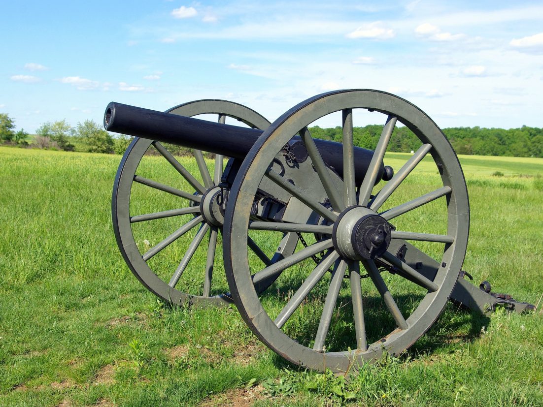
Fascinating Cemeteries – Gettysburg
In June of 1863 the Confederate Army under the Command of General Robert E. Lee pushed into Union territory. For three days 150,000 soldiers attacked Union forces in a series of battles near Gettysburg, PA. In those 3 days, 10,000 soldiers were killed, 30,000 injured and 10,000 captured or missing. 3,500 Union soldiers were buried at Gettysburg. The cemetery was dedicated in November of 1863 and President Lincoln delivered the Gettysburg Address during the ceremony.
Learn more here and plan your own visit from the Gettysburg National Military Park website.
Spirit Photography
Mary Lincoln with the ghosts of her husband Abraham Lincoln and her son Thomas “Tad” behind her. circa 1872
Charlie tells us about photography (also called ghost photography) – a type of photography whose primary goal is to capture images of ghosts and other spiritual entities. It was wildly popular during the Spiritualism movement that reached its peak from the 1840s to the 1920s. Photographers such as William Mumler and William Hope ran thriving businesses taking photos of people with their supposed dead relatives. Both were shown to be frauds, but “true believers”, such as Sir Arthur Conan Doyle, refused to accept the evidence as proof of a hoax.
Learn More:
Recipe Time
Charlie talked about Pemmican – the perfect recipe for these crazy times when you might just need to prep for the worst! Pemmican was invented by the native peoples of North America and is a concentrated mixture of fat and protein used as a nutritious food. You can get the recipe for pemmican here from Primal Survivor.
If you’d like a twist on it, our featured recipe is a hot version of pemmican which comes from a high school friend of Marianne’s that accidently texted it to her. She asked him about it, and he said it was a traditional Michigan Indian dish that would be served at funerals and festivals. Thank you, John, and Kathy, for permission to share this with our listeners.
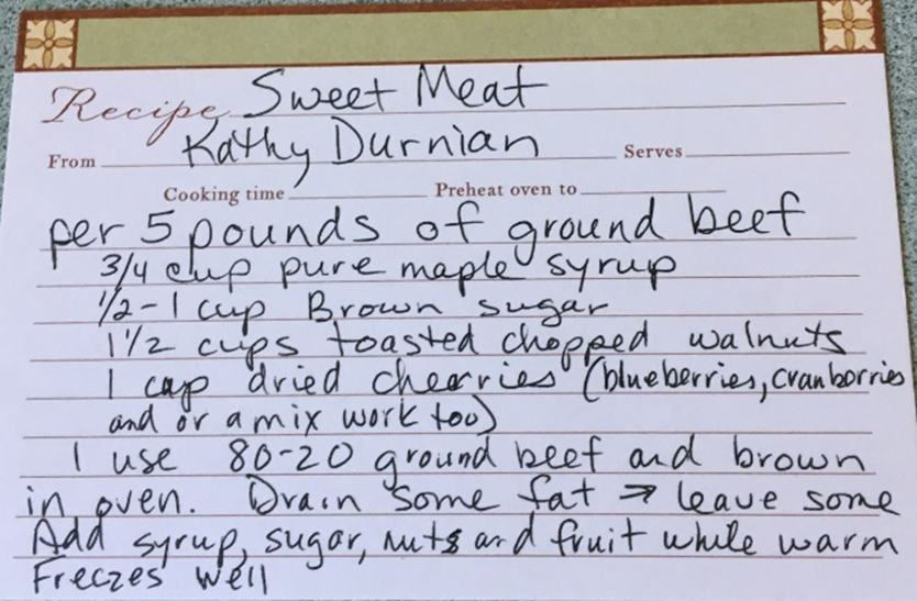
Sweet Meat
For every 5 pounds of ground beef:
- ¾ cup pure maple syrup
- ½-1 cup brown sugar
- 1 ½ cups toasted chopped walnuts
- 1 cup dried cherries (blueberries, cranberries, and/or a mix works, too)
Brown beef in oven, drain some fat, leave some. Add rest of ingredients. Serve warm over rice.